
Research
Visualizing Unexplored Spatiotemporal Scales with Synchrotron Radiation to Advance Materials Science
Many practical materials and devices possess hierarchical and heterogeneous structures extending from the atomic (microscopic) to the millimeter (macroscopic) scale, and their properties and functions are governed by diverse phenomena occurring across both space and time. To understand and design such materials, it is essential to comprehensively and quantitatively elucidate the relationships between structure and properties across scales ranging from microscopic to macroscopic and from static to dynamic. Our laboratory develops advanced synchrotron X-ray imaging and spectroscopy techniques together with sophisticated data-analysis methods to explore previously inaccessible spatiotemporal regimes spanning the nanoscale, microscale, and time domain. In particular, we combine coherent X-ray measurements with data-driven approaches to develop advanced ptychographic imaging techniques capable of visualizing dynamic phenomena that have been difficult to capture using conventional methods. Through these developments, we aim to deepen our understanding of reaction mechanisms and degradation processes in battery and catalyst materials, relationships between structure, performance, and reliability in next-generation semiconductor devices, and nucleation, growth, deformation, and fracture dynamics in metallic materials.
Major Ongoing Research Projects
| ・ |
Development of High-Spatial-Resolution Tender and Hard X-ray Ptychography Systems At NanoTerasu BL10U, we are developing a high-spatial-resolution ptychography system covering a wide photon-energy range from tender to hard X-rays. By combining advanced X-ray focusing optics with the high-speed two-dimensional CITIUS detector, we aim to achieve nanoscale imaging of a wide variety of materials. We have demonstrated broadband high-resolution ptychographic imaging and high-sensitivity measurements using CITIUS, and are pursuing spatial resolutions below 10 nm in the tender X-ray regime. 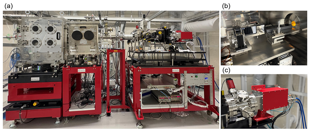
(a) Ptychography system, (b) Advanced Kirkpatrick–Baez mirrors, and (c) CITIUS detector
Related publications: ・Y. Sasaki et al., “Broadband high-resolution X-ray ptychography system spanning tender to hard X-ray regimes,” DOI IUCrJ 13, 11–18 (2026). Part of the cover image ・N. Ishiguro et al., “Towards Sub-10 nm Spatial Resolution by Tender X-ray Ptychographic Coherent Diffraction Imaging,” DOI Applied Physics Express 17, 052006 (2024). (Spotlight) (47th JSAP Outstanding Paper Award) ・Y. Takahashi et al., “High-resolution and high-sensitivity X-ray ptychographic coherent diffraction imaging using the CITIUS detector,” DOI Journal of Synchrotron Radiation 30, 989–994 (2023). Cover ・Y. Takahashi et al., “Towards High-Resolution Ptychographic X-ray Diffraction Microscopy,” DOI Physical Review B 83, 214109 (2011). |
| ・ |
Chemical-State Imaging of Battery and Catalyst Materials by X-ray Spectromicroscopy We develop X-ray spectromicroscopy techniques to visualize not only elemental distributions but also chemical states at the nano- and microscale by acquiring microscopic images while varying the incident X-ray energy across absorption edges. Using ptychography-XAFS and full-field X-ray microscopy-XAFS, we investigate spatial heterogeneity in chemical states and changes associated with reactions and degradation in battery materials, catalysts, oxygen-storage materials, and other functional materials. We have demonstrated two-dimensional, three-dimensional, and operando chemical-state imaging of lithium-ion batteries, lithium–sulfur batteries, all-solid-state batteries, and oxygen-storage materials.
Lithium–sulfur battery material: spatial distribution of S–C bonds
Lithium–sulfur battery material: spatial distribution of S–S bonds
Ce valence-state distribution in a ceria–zirconia solid-solution catalyst
・Y. Sasaki et al., “Three-dimensional imaging of sulfur chemical states in polymers with micrometer thickness using sulfur K-edge ptychographic tomography,” DOI Scientific Reports 16, 26655 (2026). ・N. Ishiguro et al., “Comprehensive Operando Visualization of the Electrochemical Events in the Cathode/Anode Layers in Thin-Film-Type All-Solid-State Lithium-Ion Batteries,” DOI ACS Applied Energy Materials 6, 8306–8315 (2023). ・M. Abe et al., “Visualization of Sulfur Chemical State of Cathode Active Materials for Lithium–Sulfur Batteries by Tender X-ray Spectroscopic Ptychography,” DOI The Journal of Physical Chemistry C 126, 14047–14057 (2022). ・H. Uematsu et al., “Visualization of Structural Heterogeneities in Particles of Lithium Nickel Manganese Oxide Cathode Materials by Ptychographic X-ray Absorption Fine Structure,” DOI The Journal of Physical Chemistry Letters 12, 5781–5788 (2021). ・M. Hirose et al., “Oxygen-diffusion-driven oxidation behavior and tracking areas visualized by X-ray spectro-ptychography with unsupervised learning,” DOI Communications Chemistry 2, 50 (2019). ・M. Hirose et al., “Visualization of Heterogeneous Oxygen Storage Behavior in Platinum-Supported Cerium-Zirconium Oxide Three-Way Catalyst Particles by Hard X-ray Spectro-Ptychography,” DOI Angewandte Chemie 130, 1490–1495 (2018). Frontispiece |
| ・ |
Development of Dynamic Coherent X-ray Diffraction Imaging Using a Triangular Aperture and Its Application to Microstructural Dynamics in Metallic Materials We are developing dynamic coherent X-ray diffraction imaging using a triangular aperture, which enables sample images to be reconstructed from a single diffraction pattern without raster scanning. We have demonstrated single-frame imaging of extended objects and, by combining the method with X-ray photon correlation spectroscopy (XPCS), achieved particle-motion analysis over length scales ranging from nanometers to micrometers. We are applying these techniques to dynamic processes in metallic materials, including nucleation, growth, deformation, and fracture.
Dynamic imaging of Brownian motion of gold colloidal particles
Dynamic imaging of microstructure formation in an LPSO-type Mg alloy during heating
・S. Takazawa et al., “Spatiotemporal Mapping of Alloy Mesostructure Dynamics via Multimodal Coherent X-ray Diffraction Imaging,” DOI Proceedings of the National Academy of Sciences of the United States of America 122, e2513369122 (2025). ・S. Takazawa et al., “Coupling X-ray Photon Correlation Spectroscopy and Dynamic Coherent X-ray Diffraction Imaging: Particle Motion Analysis from Nano-to-Micrometer Scale,” DOI Physical Review Research 5, L042019 (2023). ・S. Takazawa et al., “Demonstration of single-frame coherent X-ray diffraction imaging using triangular aperture: Towards dynamic nanoimaging of extended objects,” DOI Optics Express 29, 14394–14402 (2021). ・J. Kang et al., “Single-frame coherent diffraction imaging of extended objects using triangular aperture,” DOI Optics Express 29, 1441–1453 (2021). |
| ・ |
Advancement of X-ray Ptychography and Ptychographic CT for Semiconductor Materials and Device Analysis We have developed multislice X-ray ptychography to achieve high-spatial-resolution imaging of thick specimens by taking X-ray propagation within the specimen into account. Our previous studies established multislice reconstruction in which a thick specimen is represented by multiple slices, precession X-ray ptychography using measurements at different sample orientations, and three-dimensional reconstruction methods based on the multislice approach. More recently, we have developed a ptychographic CT system at NanoTerasu by combining tender X-ray ptychography with high-precision sample rotation. Taking advantage of the high sensitivity of tender X-rays to light elements together with their appropriate penetration depth, we have demonstrated three-dimensional discrimination between Si and SiO2 inside CMOS image sensors and nondestructive three-dimensional visualization of high-aspect-ratio etched holes for 3D NAND flash memory. We are currently extending ptychographic CT by incorporating the multislice approach to enable more accurate and higher-resolution three-dimensional imaging of thick specimens while accounting for X-ray propagation and multiple-scattering effects.
Three-dimensional reconstructed image of the internal structure of a CMOS image sensor
Three-dimensional reconstructed image of high-aspect-ratio etched holes for 3D NAND flash memory
・N. Okawa et al., “Nondestructive structural evaluation of high-aspect-ratio-etched holes by tender X-ray ptychography with computed tomography,” DOI Applied Physics Letters 129, 011109 (2026). ・N. Okawa et al., “Three-dimensional imaging of CMOS image sensor pixel structures using ptychographic X-ray computed tomography in the tender X-ray regime,” DOI Applied Physics Letters 127, 184102 (2025). Cover Picture (Featured Article) ・K. Shimomura et al., “Three-dimensional iterative multislice reconstruction for ptychographic X-ray computed tomography,” DOI Optics Express 26, 31199–31208 (2018). ・K. Shimomura et al., “Precession X-ray Ptychography with Multislice Approach,” DOI Physical Review B 91, 214114 (2015). ・A. Suzuki et al., “High-resolution multislice x-ray ptychography of extended thick objects,” DOI Physical Review Letters 112, 053903 (2014). |
| ・ |
Development of Data-Driven X-ray Imaging and Real-Time Analysis Methods We develop data-driven methods for the rapid and high-accuracy reconstruction and analysis of large-scale diffraction datasets acquired by coherent X-ray imaging. We have demonstrated amplitude reconstruction from single-frame diffraction data using image filtering, as well as improved ptychographic and X-ray fluorescence imaging based on virtual single-pixel imaging. We are also developing novel imaging approaches, including X-ray ghost imaging using structured illumination and intensity correlations. Furthermore, by incorporating physical laws and measurement principles into deep learning, we have achieved fast and accurate image reconstruction for dynamic coherent X-ray diffraction imaging. We are currently developing systems for real-time image reconstruction and analysis using machine learning during synchrotron radiation experiments. Related publications: ・T.-S. Vu et al., “PID3Net: A Deep Learning Approach for Single-Shot Coherent X-ray Diffraction Imaging of Dynamic Phenomena,” DOI npj Computational Materials 11, 66 (2025). ・M. Abe et al., “Guided image-filtering-assisted phase retrieval for amplitude reconstruction in single-frame coherent diffraction imaging,” DOI Optica 11 (2024). Cover ・M. Abe et al., “X-ray ptychographic and fluorescence microscopy using virtual single-pixel imaging based deconvolution with accurate probe images,” DOI Optics Express 31, 26027–26039 (2023). |
| ・ |
Development of X-ray Spectromicroscopy, High-Speed XAFS Measurement, and Data-Analysis Methods We develop measurement and data-analysis methods based on X-ray absorption fine structure (XAFS) for high-spatial-resolution and high-efficiency characterization of local structures and chemical states in materials. Our research includes nanoscale determination of interatomic distances by combining spectro-ptychography with EXAFS, phase-retrieval methods utilizing the Kramers–Kronig relation, restoration of XAFS spectra from sparsely sampled spectroscopic ptychography data, and high-speed XAFS measurement systems for synchrotron beamlines. Related publications: ・N. Ishiguro et al., “XAFS measurement system using undulator synchronized on-the-fly monochromator scan at SPring-8 BL36XU,” DOI Journal of Synchrotron Radiation 33 (2026). ・N. Ishiguro and Y. Takahashi, “Method for restoration of X-ray absorption fine structure in sparse spectroscopic ptychography,” DOI Journal of Applied Crystallography 55, 929–943 (2022). ・M. Hirose et al., “Nanoscale determination of interatomic distance by ptychography-EXAFS method using advanced Kirkpatrick–Baez mirror focusing optics,” DOI Journal of Synchrotron Radiation 27, 455–461 (2020). ・M. Hirose et al., “Use of Kramers-Kronig relation in phase retrieval calculation in X-ray spectro-ptychography,” DOI Optics Express 25, 8593–8603 (2017). |
| ・ |
Development of Multibeam X-ray Ptychography Systems We are developing multibeam X-ray ptychography, in which multiple coherent X-ray beams illuminate a sample simultaneously, to improve the efficiency of synchrotron radiation utilization and achieve wide-field, high-resolution imaging. At SPring-8, we demonstrated multibeam ptychography using three X-ray beams for the first time and showed that a wider field of view can be imaged within the same measurement time compared with conventional single-beam ptychography. We are further developing high-efficiency and high-speed ptychography systems using larger numbers of beams, with different phase modulations applied to individual beams to improve the stability of phase-retrieval calculations. Related publications: ・M. Hirose et al., “Multibeam ptychography with synchrotron hard X-rays,” DOI Optics Express 28, 1216–1224 (2020). ・M. Ikenaga et al., “Development of phase-modulated X-ray multibeam ptychography with total reflection focusing mirrors,” DOI Optics Express 34, 35077-35088 (2026). |
| ・ |
Development of Bragg X-ray Ptychography for Imaging Crystal Strain and Defects We are developing Bragg X-ray ptychography, which combines Bragg diffraction with ptychographic imaging to visualize strain and defect structures in crystalline materials with high spatial resolution. We have demonstrated nanoscale visualization of the strain field around a dislocation in a silicon single crystal and showed the possibility of generating an X-ray vortex beam using the associated phase singularity. We are now extending this concept to energy-dispersive Bragg ptychography, in which the Bragg condition is scanned by varying the incident X-ray energy, with the aim of high-precision three-dimensional imaging of strain and crystal-structure distributions in crystalline thin films and semiconductor devices. Related publications: ・Y. Takahashi et al., “Bragg x-ray ptychography of a silicon crystal: Visualization of the dislocation strain field and the production of a vortex beam,” DOI Physical Review B 87, 121201(R) (2013). |
| ・ |
Development of Low-Dose Imaging by Dual-Beam X-ray Ptychography To achieve high-resolution imaging of radiation-sensitive materials, we are developing dual-beam X-ray ptychography using two coherent X-ray beams. By interfering a strong reference beam with a weak beam transmitted through the sample, this approach enhances sensitivity to phase information under low-photon-count conditions and aims to reconstruct images with substantially lower radiation dose than conventional methods. We are currently advancing both measurement and reconstruction methods toward a significant reduction in the required dose. |
