Research
Takata Laboratory aims to visualize the electronic structures of materials at the nanometer level using synchrotron radiation (SR) X-rays and establish the design guidelines for the development of new functional materials. We develop advanced measurement techniques using the characteristics of rapidly evolving SR light sources as well as data analysis methods. Furthermore, we apply these methods to a variety of functional materials, ranging from hard to soft matter.
Our current research topics consist of the following:
- Next generation SR facility (NanoTerasu) project
- Innovation of structural science through the development of X-ray visualization technology
- Development of soft X-ray microscopes and their application to biological samples
- Development of soft X-ray operando spectroscopies and their application to surface and interface processes
- Development of soft X-ray light sources, optical elements, and detectors.
1. Next generation SR facility (NanoTerasu) project
Currently, we are strongly promoting the “Next generation SR facility (NanoTerasu)” (Fig. 1), which is a state-of-the-art soft X-ray light source. We aim to innovate X-ray nano visualization technology by combining the unprecedented light properties of the next generation SR facility with our own X-ray diffraction/microscopy/spectroscopy techniques and data analysis methods, such as the maximum entropy method. In addition, we aim to make the next generation SR facility a cradle of open innovation through a new type of industry-academia collaboration based on the “Coalition concept”, in which stakeholders from both academia and industries form a strong one-on-one team for the purpose of solving social challenges.

2. Innovation of structural science through the development of X-ray visualization technology
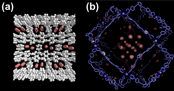
The properties and functions of materials are determined by the distribution and movement of the electrons within them. Conventional structural science has clarified the atomic arrangement of materials. We have succeeded in precisely mapping the electron density distribution and the electrostatic potential in materials by combining X-ray diffraction experiments using high-brilliance SR X-rays with the maximum entropy method. This makes it possible to visualize the interaction between the atoms and molecules in materials. So far, we have applied our X-ray nano visualization technology to various functional materials, such as metal-organic frameworks (MOFs), fullerenes, superconductors, dielectrics, multiferroics, boron compounds, and ceramics (Fig. 2).
3. Development of soft X-ray microscopes and their application to biological samples
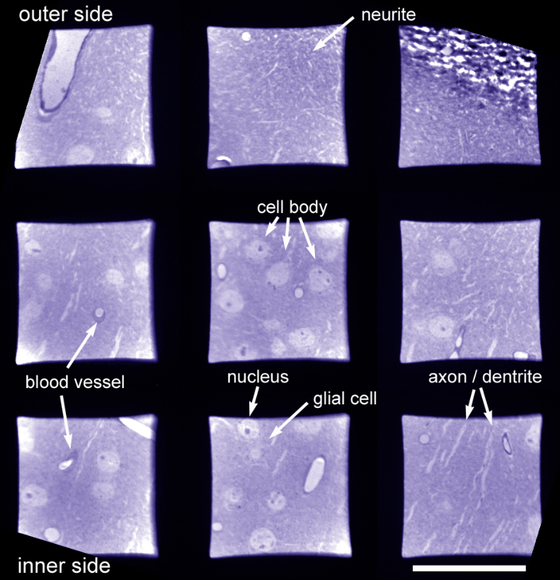
Biological samples are usually observed using optical microscopes with visible light (VIS). However, the spatial resolution of microscopes is limited by the wavelength of incident light. With soft X-ray (SX) microscopes, we can improve the spatial resolution because SX has the wavelength of one or more orders shorter than VIS. Handling SX requires special optical elements due to the short wavelength of SX and the strong interaction of SX with matter. We are now developing SX microscopes with our own technologies for SX optics and detectors. We are also applying them to biological samples (Fig. 3).
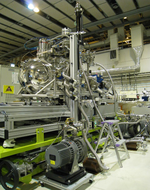
4. Development of soft X-ray operando spectroscopies and their application to surface and interface processes
“Operando measurement” is a measurement method wherein devices and heterogeneous catalysts under operation are directly monitored and their operating mechanisms are elucidated. We have developed an ambient pressure soft X-ray photoelectron spectroscopy (AP-XPS) system at SPring-8 BL07LSU (Fig. 4), in collaboration with the Institute for Solid State Physics, the University of Tokyo. This system allows operando XPS measurements in a gas atmosphere consisting of several Torr. We focus on industrially important catalytic reactions, such as CO2 hydrogenation. In addition, we are now developing instrumentation for the next generation SR facility, which is aimed at improving gas pressure and time resolution for the AP-XPS system.
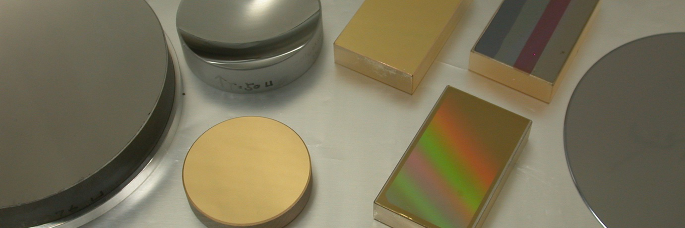
5. Development of soft X-ray light sources, optical elements, and detectors
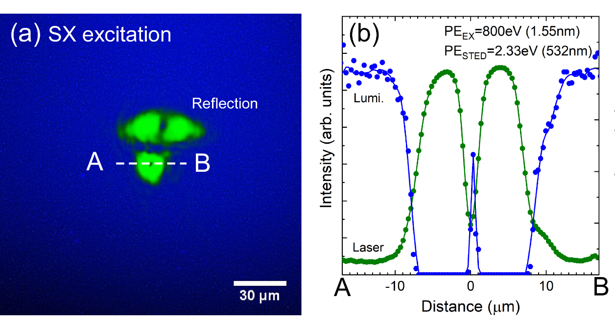
The interaction of soft X-rays with matter is much stronger than that of visible light. This makes the optical techniques in the SX region different from those in the VIS region. Short penetration depth of SX into materials (less than 1 μm) requires reflection-type optics in the SX region. Reflective mirrors and diffraction gratings are used in microscopic and spectroscopic optics, respectively, rather than transmission-type lens and prisms. Multilayer-coated optics tailored for a specific energy region can be used as high throughput (large numerical aperture and high reflectance) optics in normal incidences or in small angles of incidence geometry (Fig. 5). Grazing incidence optics can be used in a wide energy region. We are developing lateral- and depth- thickness distribution control techniques in the multilayer deposition. Light sources and detectors are also unique in the SX region because of the strong interaction of SX with matter. One of our recent developments for SX detectors is the demonstration of a stimulated emission depletion phenomenon, which will be used in SX super-resolution technology (Fig. 6).